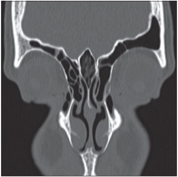
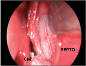
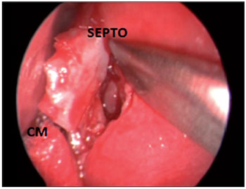
Middle ear adenoma with neuroendocrine differentiation: relate of two cases and literature review
Image was published in :2013 Vol.: 17 Issue.: 3 - 15
Description:
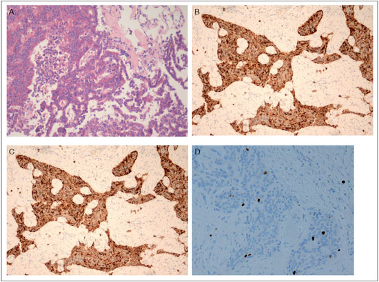
Figure 6. A: Well-differentiated papillary/trabecular/solid epithelial neoplasm composed of medium-sized cells. Central nuclei presented salt-and-pepper - like chromatin, and granular eosinophilic cytoplasm. Some neoplastic cells were seen amidst a fibrotic stroma. HE stain, original magnification x200. B-C: Immunohistochemical identification keratins marked by the antibodies AE1+AE3 and most of them were reactive for neuroendocrine markers chromogranin and synaptophysin. Original magnification x100. D: Ki-67 Ag was expressed in very low level (< 1 % of the cells). Original magnification x400.
Author(s) of the article:
Aline Gomes Bittencourt1, Robinson Koji Tsuji2, Francisco Cabral Junior3, Larissa Vilela Pereira3, Anna Carolina de Oliveira Fonseca1, Venâncio Alves4, Ricardo Ferreira Bento5.