Image was published in :2013 Vol.: 17 Issue.: 3 - 17
Description:
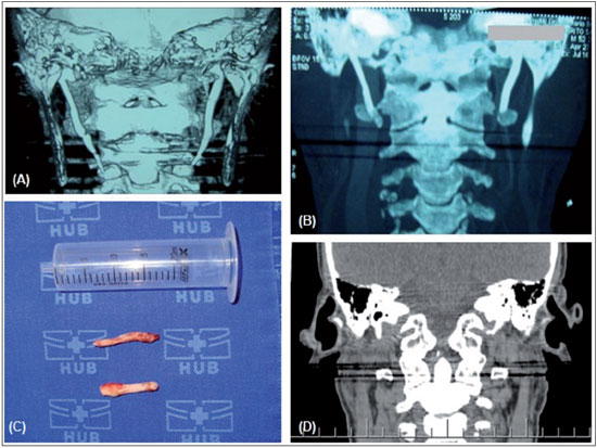
FIGURE 1. (A) Computed tomography (CT) scan of the neck showing elongation and ossification of the styloid processes of the temporal bone. The right styloid process measures 5.7 cm and the left measures 4.9 cm, which is compatible with Eagle's syndrome. (B) CT scan of the neck after intraoral styloidectomy, showing ossification of the styloid processes. The right styloid process measures 4.4 cm and the left measures 3.9 cm, thus suggesting that the base of the styloid process is present on both sides. (C) Surgical specimen after extraoral styloidectomy: right and left styloid processes. (D) CT scan of the neck after extraoral styloidectomy, showing the absence of elongation of the styloid processes on both sides.
Author(s) of the article:
Thaís Gonçalves Pinheiro1, Vítor Yamashiro Rocha Soares2, Denise Bastos Lage Ferreira3, Igor Teixeira Raymundo3, Luiz Augusto Nascimento4, Carlos Augusto Costa Pires de Oliveira5.